Protocol arterial extremity ultrasound Upper extremity arterial velocities ultrasound Ultrasound arterial doppler patient preparation
Ultrasound Assessment of Lower Extremity Arteries | Radiology Key
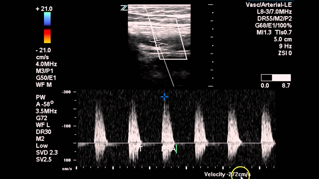
Fig. 6. adjustment of pulsed-wave doppler ultrasonography in a stenotic Duplex ultrasound technical considerations for lower extremity venous Peroneal vein ultrasound
Mobile arterial ultrasound
Computational methods to automate the initial interpretation of lowerBlood clot in leg ultrasound [diagram] lower extremities diagramLower extremity arterial ultrasound protocol manual.
Peripheral arterial ultrasound evaluationsLower extremity arterial ultrasound Venous duplex extremity lower protocol dvt sonographic suarez veins sonographictendenciesBilateral lower extremity arterial duplex.

Doppler ultrasound arterial extremity
[diagram] doppler ultrasound of left lower extremity superficial wiringDoppler ultrasound images of the lower right limb sho Lower extremity venous ultrasound worksheetLower extremity arterial doppler worksheet.
Venous duplex extremity protocol sonographictendenciesUltrasound arterial peripheral vascular exams arteries artery doppler evaluations extremity common femoral patient ankle brachial exam flow anatomy sonography cardiac Ultrasound venous extremity duplex dvt vein acute considerations thrombosis invisible grayscale virtuallyArterial ultrasound.
![[DIAGRAM] Lower Extremities Diagram - MYDIAGRAM.ONLINE](https://i2.wp.com/ultrasound.simplybook.me/uploads/ultrasound/event__picture/original/97ef512a1ac31da8dfeb6d9267434d28.png)
Assessment of upper extremity arterial disease
Upper extremity arterial ultrasound worksheetFigure 4 from doppler ultrasonography of the lower extremity arteries Pin by ramzi azzam on imagiologiaProtocol arterial extremity lower manual ultrasound pricing.
Anatomy lower extremity arteries vascular ultrasound limb system peripheral body radiology medical bing savedUltrasound vascular dvt anatomy leg sonography medical physics school radiology imaging entire student tech diagnostic board choose Protocol for performing lower extremity arterial duplexLower doppler extremity figure arteries anatomy ultrasonography scanning guidelines.

Lower extremity arterial doppler
Lower extremity venous duplex protocol – sonographic tendenciesUpper arterial extremity disease assessment radiology fig Lower extremity arteries anatomyDuplex lower arterial extremity bilateral study ultrasound vascular occlusion left case sfa disease radiology imaging.
Doppler ultrasound of lower limb arteriesUltrasound assessment of lower extremity arteries Ultrasound extremity arteries radiologyDvt extremity venous ultrasound imaging findings normal iem emergency.

Lower extremity deep venous us imaging – illustrations – international
Lower extremity venous duplex protocol – sonographic tendenciesDoppler ultrasound limb arteries arterial aorta abdominal anatomy graft Lower extremity arterial ultrasound worksheetLower extremity arterial ultrasound protocol manual.
Lower limb arteries lower limb ct angiography anatomy radiologyDoppler ultrasound of lower limb arteries Point‐of‐care ultrasound for deep venous thrombosis of the lower limbDoppler ultrasound limb arteries.

Extremity upper ultrasound arterial protocol duplex lower assessment doppler utilizing performing vascular anatomy studylib
Pdf doppler ultrasonography of the lower extremity arteries anatomy .
.

Bilateral Lower Extremity Arterial Duplex - Case Study - YouTube

Protocol for Performing Lower Extremity Arterial Duplex

Pdf Doppler Ultrasonography Of The Lower Extremity Arteries Anatomy

Lower Extremity Venous Duplex Protocol – Sonographic Tendencies

Lower Extremity Arterial Doppler Worksheet

Fig. 6. Adjustment of pulsed-wave Doppler ultrasonography in a stenotic
